
Selected presentation abstracts, with an informal introduction from the speakers.

Megapixels per minute: Chemical imaging using fast X-ray fluorescence microscopy
Australian Synchrotron, X-ray fluorescence microscopy beamline.
I will always remember acquiring my first megapixel X-ray fluorescence image in half an hour using a prototype Maia detector. My memories of that moment are sharpened by the response of my friends when I excitedly broke the news – “So what! my cheap digital camera can take 4 megapixel images in a second.”
I then explained to my friends that the same scan with old detector technology and data acquisition methods would have taken 2 weeks. What we had achieved was more than a 1000 times faster - the type of quantum leap that happens rarely in science and technology. The Maia detector and event mode data acquisition was a beautiful example of disruptive technology and paradigm shift.
That was 6 years ago, X-ray fluorescence microscopy running at megapixels per hour. Now we are pushing megapixels per minute, and still have untapped potential to couple fast XFM with even brighter diffraction limited storage rings and higher resolution, more efficient focussing optics.
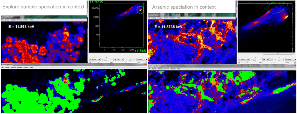
In my talk I will describe how we have harnessed the capabilities of fast XFM to enable chemical speciation imaging or fluorescence XANES imaging. XANES imaging enables researchers to investigate chemical speciation in-situ with real spatial context at environmentally relevant concentrations. Finally I will also touch on improvements to efficiency and throughput by deploying advanced motion sequencing.
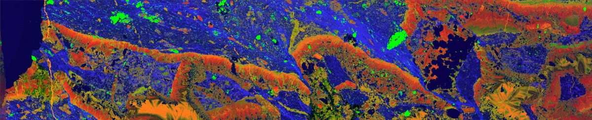
Sr (red): Fe (green): Rb (blue) Gigapixel image: 40 × 9 mm = 66667 × 1500 (600 nm) pixels, 133 μsec dwell, scan time 38 hrs . Petrographic section of high grade ore from western shear zone of the Sunrise Dam gold deposit, WA. Used in CSIRO research to understand the elemental associations of gold and its distribution in complex mineral phases. Demonstration of fast XFM capabilities and ability to generate Gigapixel 2D and 3D data.
Abstract
MEGAPIXELS PER MINUTE: CHEMICAL IMAGING WITH FAST X-RAY FLUORESCENCE MICROSCOPY
D. Paterson1, D. L. Howard1, M. D. de Jonge1, K. M. Spiers2, C. G. Ryan3, R. Kirkham3, B. E. Etschmann4, S. A.James5, E. Lombi6, E. Donner6, P. M. Kopittke7
1. Australian Synchrotron, Blackburn Road, Clayton VIC 3168, Australia
2. PETRA III, DESY, 22607 Hamburg, Germany
3. CSIRO Mineral Resources, Normanby Road, Clayton VIC 3168, Australia
4. Monash University, Clayton VIC 3168, Australia,
5. Florey Institute of Neuroscience and Mental Health, Parkville VIC 3052, Australia
6. University of South Australia, Mawson Lakes SA 5095, Australia
7. University of Queensland, St Lucia QLD 4072, Australia
X-ray fluorescence microscopy (XFM) can be used for elemental and chemical microanalysis across length scales ranging from millimeter to nanometer. XFM is ideally suited to quantitatively map trace elements within whole plant, biological specimens, environmental and soil samples. The high elemental sensitivity of the X-ray fluorescence microprobe combined with deep penetration of hard X-rays enables measurement of whole cells, tissue sections and a diverse range of environmental samples with a minimum of preparation.
Event mode X-ray fluorescence detection methods pioneered by the Maia detector [1, 2, 3] at the Australian Synchrotron XFM beamline [4] enable high definition imaging approaching megapixel per minute rates. The ability to rapidly acquire 2D images enables higher-dimensional studies such as fluorescence tomography, XANES imaging, and XANES tomography in realistic times. Fast event mode XFM has enabled the realisation of extremely-fractionated x-ray fluorescence tomography [5].
Full spectral XANES imaging takes advantage of fast XFM and results in X-ray absorption near edge structure spectra from X-ray fluorescence at each pixel in an image. The technique was first demonstrated in an inhomogeneous geomaterial an oxidized pisolitic regolith [6]. Recently the technique has been described as fluorescence imaging XANES (ϕXANES) and used to visualize in vivo coordination environments of metals in biological specimens [7].
ϕXANES has been employed with micron resolution and moderate definition (10K pixels, 100–200 energies) across a diverse range of sciences and applications from environmental chemistry [8] to arsenic toxicity in crop production [9]. Studies probing and optimising the efficiency and sensitivity of ϕXANES to achieve robust measurements at environmentally relevant concentrations will be presented. The major remaining constraint to higher definition or faster more efficient imaging is the line overheads in scan motion. A motion sequencing upgrade being deployed to address this restriction will be discussed.
References
- D. P. Siddons et al., AIP Conference Proceedings 705, 953 (2004).
- R. Kirkham et al., AIP Conference Proceedings 1234, 240 (2010).
- D. P. Siddons et al., J. of Physics: Conf. Series 499, 012001 (2014).
- D. Paterson et al., AIP Conference Proceedings 1365, 219 (2011).
- M. D. de Jonge et al., XRM2016 Abstracts.
- B. E. Etschmann et al., American Mineralogist 95, 884 (2010).
- S. A. James et al., Scientific Reports 6, 20350 (2016).
- B. E. Etschmann et al., Environmental Chemistry, 11, 341 (2014).
- P. M. Kopittke, et al., New Phytologist 201, 1251 (2014).
David Paterson
 David Paterson led the development of the Australian Synchrotron’s X-ray Fluorescence Microscopy beamline and has more than two decades of experience in synchrotron radiation research.
He has a passionate interest in using X-ray fluorescence microscopy to improve our lives particularly in the fields of environmental science and new materials research.
His research interests are focused on disruptive technology in X-ray fluorescence microscopy, particularly detector and data acquisition schemes. He has fostered and promoted the R&D100 awarded Maia detector at the Australian Synchrotron.
David enjoys skiing, bike riding and playing bass in a garage jazz band.

Diamond Light Source is the UK's national synchrotron science facility, located at the Harwell Science and Innovation Campus in Oxfordshire.
Copyright © 2022 Diamond Light Source
Diamond Light Source® and the Diamond logo are registered trademarks of Diamond Light Source Ltd
Registered in England and Wales at Diamond House, Harwell Science and Innovation Campus, Didcot, Oxfordshire, OX11 0DE, United Kingdom. Company number: 4375679. VAT number: 287 461 957. Economic Operators Registration and Identification (EORI) number: GB287461957003.