
Selected presentation abstracts, with an informal introduction from the speakers.

Multiple-wavelength resonant fluctuation x-ray scattering
European XFEL GmbH, theory and simulations.
One of the most attractive challenges in x-ray free electron laser (XFEL) science is to image an individual particle, for example a virus, at high resolution. This could enable structural studies of the nanoscience and biology of those materials—such as understanding their properties and functions—that cannot be accessed by conventional means, for example x-ray crystallography. In a typical single-particle imaging experiment, individual particles are injected in to the x-ray beam to produce diffraction patterns on a detector before their destruction by intense X-radiation. These diffraction patterns encode structural information about the particle. Importantly, structural changes in a material can be detected on femtosecond timescales at an XFEL, due to its ultrashort, ultrabright coherent pulses. Therefore, not only the static features, but also the dynamical behavior of particles can be studied.
Even at such high-brilliance x-ray sources as an XFEL, it is a challenging task to detect enough scattered photons from a small radiation-sensitive particle. A concept “scatter from many—understand the single” offers an alternative way for single-particle structure determination, which is accomplished by scattering from a disordered ensemble of many reproducible particles rather than just one. Clearly, larger numbers of objects in the x-ray beam produce higher scattered intensities, giving another opportunity to recover the structure of a single object. The puzzle to solve is how to obtain single-particle features from the x-ray signal recorded from an ensemble of many randomly distributed particles. Conventional approaches relying on the regular structure of crystalline objects cannot be applied due to a lack of translational symmetry in a disordered system of particles. It appears that by applying angular intensity cross-correlation functions to the scattered signal from many particles it is still possible to extract single-particle information.
In the Theory group of the European XFEL in Hamburg (Germany) we are working on the development of these cross-correlation based approaches for single-particle structure determination. Recently developed methods have, particularly, been applied to recover the structure of nanosized viruses from the experimental data measured at LCLS, an XFEL in Stanford (USA), in a series of experiments carried out by a large international Single Particle Imaging (SPI) initiative. It is expected that XFELs with their cutting edge capabilities will open new horizons in the studies of structure and dynamics of materials.
Abstract
MULTIPLE-WAVELENGTH RESONANT FLUCTUATION X-RAY SCATTERING
R. P. Kurta
European XFEL GmbH, Albert-Einstein-Ring 19, D-22761 Hamburg, Germany
Fluctuation x-ray scattering (FXS) proposes to measure cross-correlation functions from scattered intensity fluctuations to facilitate structure determination of a single particle in solution [1]. Due to its intrinsic capability to treat multiple-particle scattering data, the FXS approach is advantageous for structural determination of weakly-scattering objects, or particles that cannot be crystallized [1-5]. With the emergence of x-ray free electron lasers, the FXS approach offers an alternative technique for single-particle structure determination in coherent diffractive imaging experiments [4, 5].
Here a multiple-wavelength resonant FXS approach is presented for element–specific imaging of nanoscale objects in random ensembles with short positional and rotational relaxation times [6]. It is shown, that by applying x-ray cross-correlation analysis in combination with iterative phase retrieval to the scattering data measured at multiple x-ray energies near an absorption edge of a substance, it is possible to image the nanoscale structure of an individual object with chemical sensitivity (Figure 1). The elemental distribution in distinct two-component model nanostructures has been reconstructed using the simulated scattering data from two-dimensional random ensembles of particles.
The CLS cryo-STXM is a component of a CFI Leading Edge fund project, "Enhancing the Spectro-microscopy Beamline and Endstations at the Canadian Light Source” (U. Sask; S.G. Urquhart, Project Leader), with partial financial support from AFCC Automotive Fuel Cell Cooperation Corp. CLS is supported by the Canada Foundation for Innovation, Natural Sciences and Engineering Research Council of Canada, the University of Saskatchewan, the Government of Saskatchewan, Western Economic Diversification Canada, the National Research Council Canada, and the Canadian Institutes of Health Research.
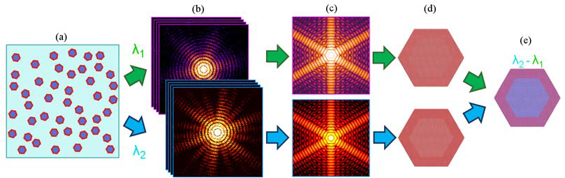
Figure 1: Outline of the multiple-wavelength resonant fluctuation x-ray scattering approach [6]. In the experiment x-ray scattering is measured from a sample composed of identical objects (a) at two different wavelengths, λ. The resulting two sets of diffraction patterns (b) are used to recover the scattered intensity distributions (c) corresponding to a single object. The recovered intensities (c) are inverted by the iterative phase retrieval technique to reconstruct the images of the object (d) measured at two distinct energies, and to determine the elemental distribution in the object (e).
References
- Z. Kam, Macromolecules 10, 927 (1977).
- M. Altarelli, R. P. Kurta, I. A. Vartanyants, Phys. Rev. B 82 (2010).
- R. P. Kurta and M. Altarelli and I. A. Vartanyants, Adv. Cond. Matt. Phys. 2013, 959835 (2013).
- B. Pedrini et al, Nat. Comm. 4, 1647 (2013).
- R. P. Kurta and R. Dronyak and M. Altarelli and E. Weckert and I. A. Vartanyants, New. J. Phys. 15, 013059 (2013).
- R.P. Kurta, J. Phys. B: At. Mol. Opt. Phys. 49, 165001 (2016)
Ruslan P. Kurta
 Theoretical Physicist at the European XFEL. X-ray scattering studies of the structure of nanoparticles and non-crystalline materials

Diamond Light Source is the UK's national synchrotron science facility, located at the Harwell Science and Innovation Campus in Oxfordshire.
Copyright © 2022 Diamond Light Source
Diamond Light Source® and the Diamond logo are registered trademarks of Diamond Light Source Ltd
Registered in England and Wales at Diamond House, Harwell Science and Innovation Campus, Didcot, Oxfordshire, OX11 0DE, United Kingdom. Company number: 4375679. VAT number: 287 461 957. Economic Operators Registration and Identification (EORI) number: GB287461957003.